3D printing, an alternative to the cadaver? Our 7 answers

April 2021
The theoretical training of medical students is supplemented by internships and practical exercises. Some of these involve carrying out examinations on cadavers in laboratories or hospitals.
Although the word may be frightening and the context may raise certain ethical questions, the fact remains that this training is absolutely essential for the proper training of medical staff, as this practice has several advantages. The main one is to allow the learning of gestures in real conditions, gestures that will never be performed the first time on the patient. The sensations allow the student surgeon to train without risk and without stress.
Nevertheless, it is clear that this type of examination has many constraints. Therefore, some alternatives have been emerging for several years to complement cadaveric examinations. One of these alternatives is medical 3D printing!
Starting from 7 identified constraints, we propose, through this article, to understand how 3D printing can be a complementary tool to cadaveric examination and bring you, we hope, some food for thought.
1 – The shortage of cadavers
One of the first constraints that appears when a university wishes to carry out cadaveric examinations is the availability of cadavers. Indeed, whether they are linked to a sensitive ethical context, to a decrease in the number of cadavers accepted due to the COVID19 pandemic or to a low number of donations due to the cost of transport often borne by families, the constraints of cadaver availability are a real scourge for student training. Finally, according to France Adot, each year about 2,500 bodies are donated to science.
3D printing makes it possible to respond to this shortage in an agile way.
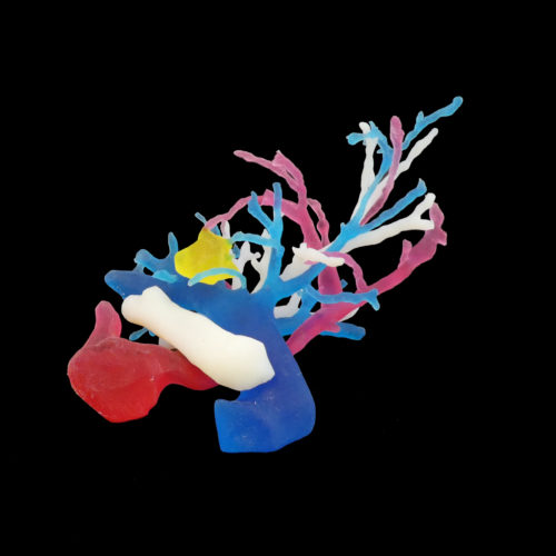
Indeed, the new technologies and materials linked to 3D printing make it possible to create anatomical models that reproduce anatomical structures in a very realistic way, both in terms of sensory feedback and material resistance, and this in record time.
2 – The absence of pathologies on cadavers
University hospitals and simulation centres train their students in a variety of specialities and the pathologies encountered are numerous. However, in addition to the lack of sufficient bodies available, it is also rare to be able to obtain a cadaver with these same pathologies in order to carry out concrete and targeted exercises.
The mantra “never the first time on the patient” can therefore seem difficult to respect.
This is where 3D printing comes in very handy: being able to recreate a specific pathology at a given location in the form of a surgical simulator. Thus, a teacher can choose the level of difficulty by opting for this or that pathology with this or that specificity. For example, it is possible to reproduce a pituitary tumour or a hand fracture… Whatever the pathology, it can be reproduced on demand in materials that perfectly represent reality. It is even possible to create several procedures to be performed on the same simulator, or to create a model based on a specific patient image.
3 – Specific and expensive logistics
The purchase and sale of cadavers is prohibited by law. Moreover, not all university structures have the necessary infrastructure to host an anatomy laboratory. A human or animal cadaver cannot simply be moved. The examinations must be carried out within a strict framework, in a dedicated space. This leads to very strong location constraints.
Conversely, 3D printed elements, whether we are talking about a surgical simulator or an anatomical model, often represent a lower overall cost. It does not require any particular storage space. Therefore, a simple storage space is sufficient, which makes things much easier without reducing the quality of the training. The organisation of practical work is thus facilitated and a classroom can thus become a mini simulation centre. It’s a real convenience!
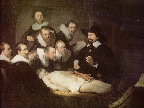
The Anatomy Lesson of Professor Tulp, Rembrandt, 1632
4 – The constraints of limited training
The previous constraints, such as the lack of available bodies, the scarcity of pathological cases or the logistical complexity, do not allow all students to train on an equal scale. Indeed, we often see practical work during which only a handful of students can practice, the others being there only for observation. Moreover, as the cadaver is “destructive”, most of the procedures cannot be performed several times. Only a few lucky ones will be able to test their techniques or experience the real thing.
Thanks to the ease of organisation and logistics made possible by 3D printed anatomical models and surgical simulators, this problem no longer exists. Each student has his own model and everyone can train at the same time. Beyond the sensations and the realization, this also allows for easier exchange between several practitioners.
This method therefore allows for greater equity in the quality of training, which is not a negligible argument when we know that universities are increasingly seeking to standardise their examinations. A simulator that is the same for everyone: this is totally in line with this (good) direction.
5- Tissue deterioration
An element that should be underlined and that is part of the conservation issues is the deterioration of cadaveric tissues. Indeed, if the training on a human body is absolutely necessary in terms of sensations, it is necessary to take into account that tissues are materials that necrose quickly. They become more friable in the course of time and no longer correspond to the reality of a living human body. This can become problematic in certain specialties such as orthopaedic surgery for example.
Once again, the answer provided by 3D printing is clear: the vast majority of materials are resistant over time and allow quality training at any time.
6 – Ethical issues related to cadaveric examinations
The use of cadaveric training, despite strict regulations, still raises ethical issues. In addition to the reluctance of families to donate the body of a loved one, healthcare personnel are also increasingly uncomfortable with these practices.
By using inanimate 3D printed material, these ethical questions no longer arise. Only the question of ecological impact may arise, as the materials used in 3D printing are not all biodegradable. However, local “on-demand” production reduces this impact thanks to the absence of unnecessary production and a greatly reduced carbon impact due to transport.
7 – Practices adapted to each academic pathway
Finally, some health professionals, such as physiotherapists, osteopaths or chiropodists, are not meant to work on cadavers or internal body parts. And yet, they need to be trained as close to reality as possible. The use of 3D printing makes it possible to meet their needs, without them having to endure too much recurrent practice on cadavers. The cadaveric examination cannot be totally replaced by alternative solutions.
However, the use of surgical simulators or 3D printed anatomical models can provide a real complement to training. The need to train on cadavers is real insofar as it will be part of their daily life. Nevertheless, complementing their training with other tools can only improve training, develop skills and offer new areas for improvement. Moreover, by confronting students with this new type of tool, they will be better able, in their professional career, to use it and to propose innovations to improve the care pathway. This means investing now in the future of medicine and care providers in general.
BONE 3D can help you in the design, development and 3D printing of medical equipment. Our team is at your disposal, contact us!